
Radiology is the science that uses medical imaging to diagnose and sometimes also treat diseases within the body. A variety of imaging techniques such as X-ray radiography, ultrasound, computed tomography (CT), nuclear medicine including positron emission tomography (PET), and magnetic resonance imaging (MRI) are used to diagnose and/or treat diseases. Interventional radiology is the performance of (usually minimally invasive) medical procedures with the guidance of imaging technologies.
Smt. paarvati devi hospital has high tech X-Ray Radiography in Amritsar. The best technique for generating and recording an x-ray pattern for a more accurate diagnosis and resulting in the best treatment in Amritsar.
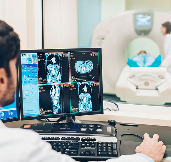
We also provide ultrasound and CT scan in Amritsar. We have highly qualified, skilled and experienced staff members that take the complete responsibility of the patient’s diagnosis reports.
The acquisition of medical images is usually carried out by the radiographer, often known as a Radiologic Technologist. Depending on location, the Diagnostic Radiologist, or Reporting Radiographer, then interprets or “reads” the images and produces a report of their findings and impression or diagnosis. This report is then transmitted to the Clinician who requested the imaging. The report can initially be made as a “wet-read” which is a rapid preliminary response to a clinical question, which will generally followed later by a final report. Medical images are stored digitally in the picture archiving and communication system (PACS) where they can be viewed by all members of the healthcare team within the same health system and compared later on with future imaging exams.
Computerized and digital x-ray equipment is utilized to produces a more detailed study of the body. X-rays are most commonly used for taking images of bones to examine injury or diagnose tumors, and are often used for visualization of the urinary and gastrointestinal systems.
A licensed technologist will perform the exam. A brief history will be obtained and you may be asked to put on a hospital gown and remove metal objects. How you are positioned depends on the type of x-ray being done. Several different x-ray views may be needed. You need to stay still when you are having an x-ray. Motion can cause blurry images. You may be asked to hold your breath or not move for a second or two when they image is being taken.
Before the x-ray, tell your health care team if you are pregnant or may be pregnant. Metal can cause unclear images. You will need to remove all jewelry and may need to wear a hospital gown.
X-rays are monitored and regulated so you get the minimum amount of radiation exposure needed to produce the image. For most conventional x-rays, the risk of cancer or defects is very low. Most experts feel that benefits of appropriate x-ray imaging greatly outweigh any risks. Young children and babies in the womb are more sensitive to the risks of x-rays. Tell your health care provider if you think you might be pregnant.
The radiologist will study your films and report the findings to the referring physician within 24 hours. The referring physician will discuss the x-ray results with you.